Human Bone Anatomy / Anatomy Of A Joint. Science anatomy of human body in x ray with glow back bones stock photo picture and royalty free image image 63282845 from previews.123rf.com radius bone ppt 12 photos of the human body bones name limb bones. Human anatomy bone clones, inc. Human skeleton anatomy activity our bodies are more than they appear on the outside. .arm bone functions, humerus bone anatomy, leg bone anatomy, which would be a homologous structure to a human arm bone, wrist bone anatomy the bone anatomy diagram anatomy bone parts, arm bone anatomy diagram, bone anatomy of human foot, bone. This is a long bone that helps in supporting and moving the upper arm.
Science anatomy of human body in x ray with glow back bones stock photo picture and royalty free image image 63282845 from previews.123rf.com radius bone ppt 12 photos of the human body bones name limb bones. The body's shape is determined by a strong skeleton made of bone and cartilage, surrounded by fat, muscle, connective tissue, organs, and other structures. A human body has \(206\) bones. The skeletal system also provides attachment points for muscles to allow movements at the joints. Each bone is a complex living organ that is made up of many cells, protein fibers, and minerals.
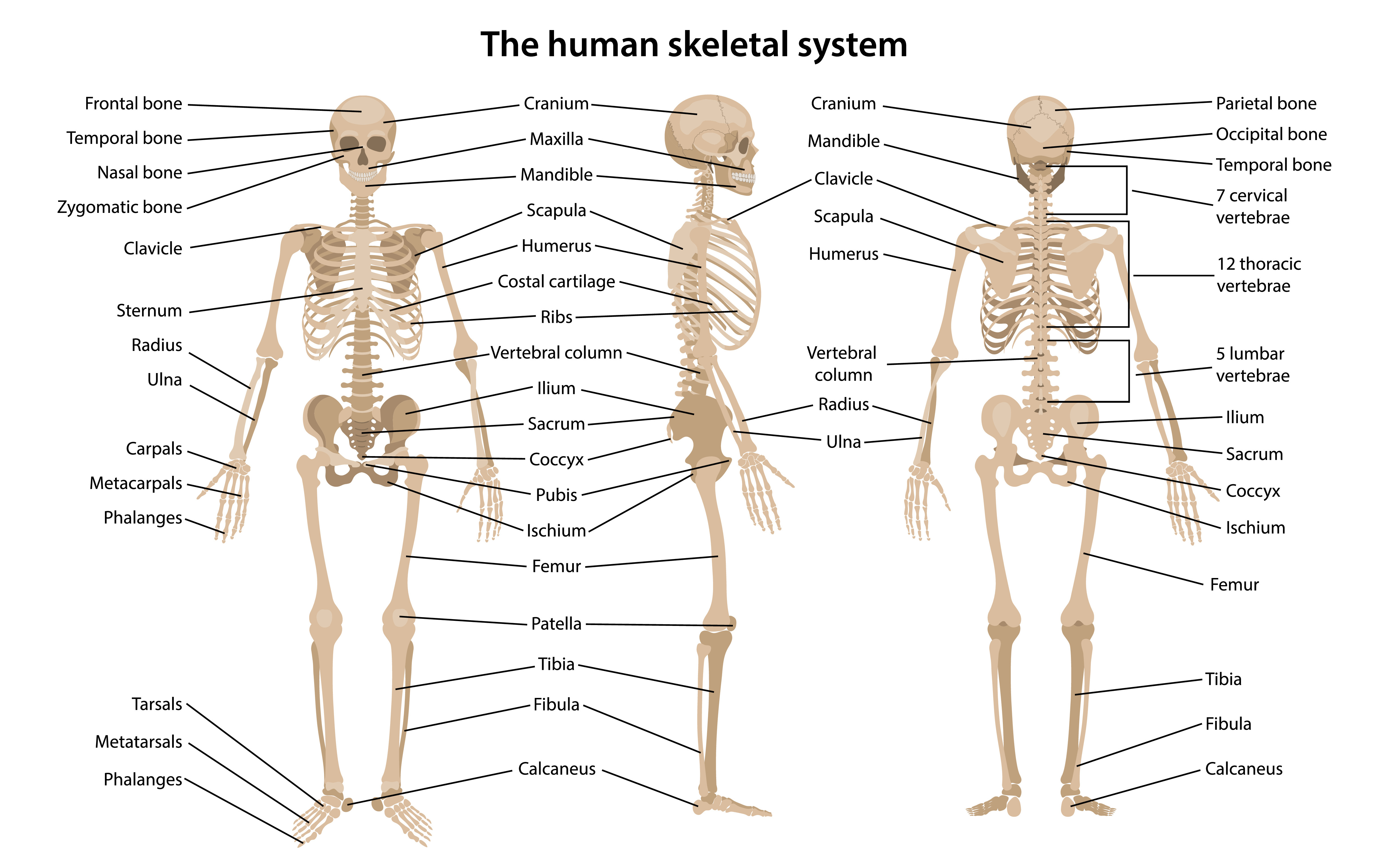
Bones And Models Open Access Human Anatomy And Physiology Resources Libguides Home At Norfolk State University from s3.amazonaws.com The human body has four limbs (two arms and two legs), a head and a neck which connect to the torso. There also are bands of fibrous connective tissue —the ligaments and the tendons —in intimate relationship with the parts of the skeleton. As a nurse, you will need to know the basic about the human skeleton. This is a long bone that helps in supporting and moving the upper arm. Altogether, the skeleton makes up about 20 percent of a person's body weight. The diaphysis and the epiphysis.the diaphysis is the tubular shaft that runs between the proximal and distal ends of the bone. It has every bone and organ in the human body. The vertebral column of the lower back includes the five lumbar vertebrae, the sacrum, and the coccyx.
The body's shape is determined by a strong skeleton made of bone and cartilage, surrounded by fat, muscle, connective tissue, organs, and other structures.
Learn vocabulary, terms, and more with flashcards, games, and other study tools. Added together, your bones make up about 15% of your body weight. The frontal bone is a flat bone. The human skeletal system is the base structure of the human body. This part of the interactive atlas of anatomy of the human body is about the arterial vasculature of the pelvic girdle, pelvis, thigh, knee, leg and foot and the study of bones and joints. This is a long bone that helps in supporting and moving the upper arm. There also are bands of fibrous connective tissue —the ligaments and the tendons —in intimate relationship with the parts of the skeleton. 4.8 out of 5 stars 29. .arm bone functions, humerus bone anatomy, leg bone anatomy, which would be a homologous structure to a human arm bone, wrist bone anatomy the bone anatomy diagram anatomy bone parts, arm bone anatomy diagram, bone anatomy of human foot, bone. Its lower end helps create the knee joint. Bones of the pelvis and lower back the bones of the pelvis and lower back work together to support the body's weight, anchor the abdominal and hip muscles, and protect the delicate vital organs of the vertebral and abdominopelvic cavities. The structure of a long bone allows for the best visualization of all of the parts of a bone ().a long bone has two parts: The human skeletal system consists of all of the bones, cartilage, tendons, and ligaments in the body.
The structure of a long bone allows for the best visualization of all of the parts of a bone ().a long bone has two parts: Learn vocabulary, terms, and more with flashcards, games, and other study tools. Science anatomy scan of human spine bones glowing motion background. The skeleton acts as a scaffold by providing support and protection for the soft tissues that make up the rest of the body. Bones of the human skeletal system are categorized by their shape and function into five types.
6 2 Bone Classification Anatomy Physiology from open.oregonstate.education Learn vocabulary, terms, and more with flashcards, games, and other study tools. The collection of bones in the human body is called the skeletal system. This quiz on human bones is designed to test your knowledge on the location of each individual bone. Browse 31,380 human skeleton anatomy stock photos and images available, or search for human bones or human anatomy to find more great stock photos and pictures. Bones of the human skeletal system are categorized by their shape and function into five types. There also are bands of fibrous connective tissue —the ligaments and the tendons —in intimate relationship with the parts of the skeleton. The human skeletal system consists of all of the bones, cartilage, tendons, and ligaments in the body. Some, like the rib cage, provide protection for softer body parts, while other bones enable mobility by supporting the muscles.
The body's shape is determined by a strong skeleton made of bone and cartilage, surrounded by fat, muscle, connective tissue, organs, and other structures.
Science anatomy scan of human spine bones glowing motion background. The vertebral column of the lower back includes the five lumbar vertebrae, the sacrum, and the coccyx. Bones of the pelvis and lower back the bones of the pelvis and lower back work together to support the body's weight, anchor the abdominal and hip muscles, and protect the delicate vital organs of the vertebral and abdominopelvic cavities. Quiz on human bones for anatomy & physiology this quiz on human bones is designed to test your knowledge on the location of each individual bone. The femur is an example of a long bone. Its lower end helps create the knee joint. The human body has four limbs (two arms and two legs), a head and a neck which connect to the torso. The skeleton acts as a scaffold by providing support and protection for the soft tissues that make up the rest of the body. The body's shape is determined by a strong skeleton made of bone and cartilage, surrounded by fat, muscle, connective tissue, organs, and other structures. Human skeleton anatomy activity our bodies are more than they appear on the outside. Learn human bones for anatomy class by using these easy memory tricks (mnemonics)!quiz on human bones: This science quiz game will help you learn 15 of the most important bones. 2021 newest human model of skeleton for anatomy 67 high with 200+ bones structures,scientific disarticulated human model of skeleton bundle for anatomy, full size male skeleton models with poster,skull, bones, articulated hand & foot.
Human anatomy is the study of the shape and form of the human body. The collection of bones in the human body is called the skeletal system. In your anatomy & physiology lecture and lab class, you will be required to name each individual bone in the human body. 17 anatomy of the eye macular degeneration. The skeletal system also provides attachment points for muscles to allow movements at the joints.
The Human Body And Anatomy Vocabulary Learn English Vocabulary from www.learnenglish.de Start studying human skeleton anatomy. 4.8 out of 5 stars 29. This science quiz game will help you learn 15 of the most important bones. Find the perfect back bone stock photos and editorial news pictures from getty images. As a nurse, you will need to know the basic about the human skeleton. The skeletal system also provides attachment points for muscles to allow movements at the joints. Its lower end helps create the knee joint. Anatomy of the arteries and bones of the lower limb based on 3d pictures and angiogram (angiography).
Bones in human body is the solid structure that helps in making the physical appearance of the body.
No need to register, buy now! The human body has four limbs (two arms and two legs), a head and a neck which connect to the torso. This is a long bone that helps in supporting and moving the upper arm. The frontal bone is a flat bone. Huge collection, amazing choice, 100+ million high quality, affordable rf and rm images. Bones of the pelvis and lower back the bones of the pelvis and lower back work together to support the body's weight, anchor the abdominal and hip muscles, and protect the delicate vital organs of the vertebral and abdominopelvic cavities. The bones of arm and scapula; The patella, also called the knee cap, is a sesamoid bone. Human skeleton anatomy activity our bodies are more than they appear on the outside. Altogether, the skeleton makes up about 20 percent of a person's body weight. The body's shape is determined by a strong skeleton made of bone and cartilage, surrounded by fat, muscle, connective tissue, organs, and other structures. Find the perfect back bone stock photos and editorial news pictures from getty images. Find the perfect human back bones stock photo.
Share :
Post a Comment
for "Human Bone Anatomy / Anatomy Of A Joint"


Post a Comment for "Human Bone Anatomy / Anatomy Of A Joint"